

What Chiropractors Miss on Imaging — and How to Sharpen Your Diagnostic Eye
Written by the ChiroCredit Education Team
Reviewed by Gregory Priest, DC, DIANM
There’s a finding on nearly every film that a rushed read walks right past — not because the doctor isn’t careful, but because most chiropractic radiology training is a fraction of what a board-certified radiologist receives. That gap doesn’t show up often. When it does, it tends to show up on exactly the cases where missing it matters most.
The Training Gap Most DCs Don’t Think About
Chiropractic education includes plain film interpretation, but it’s nowhere near the depth of a residency-trained radiologist’s education. The credential that closes that gap in the chiropractic profession is the Diplomate of the American Chiropractic Board of Radiology (DACBR) — a board-certified chiropractic radiologist who has completed years of additional, specialized training in imaging interpretation. Most practicing DCs will never pursue the DACBR itself, but learning directly from those who have is the most realistic way to narrow the gap.
Where the Misses Tend to Cluster
A few categories account for a disproportionate share of missed or delayed findings on plain film. Distinguishing a benign bone lesion from an aggressive one — or a tumor-like condition from an actual tumor — requires pattern recognition that isn’t intuitive without specific training, which is why entire courses in ChiroCredit’s imaging library are dedicated to benign versus malignant neoplasia specifically.
Arthritides present a similar problem. Differentiating osteoarthritis from inflammatory arthritis, or identifying the specific arthritide at play, changes the treatment and referral pathway entirely, and the radiographic differences are subtler than most DCs are taught to look for. Age adds another layer of difficulty: a finding that’s a normal variant in a pediatric patient can look pathological out of context, while in geriatric patients, degenerative overlap can mask a fracture or obscure a more serious finding. Reading the same film differently depending on the patient in front of you is a skill, not a default.
Chest findings round out the list. Chiropractors order and review chest films more often than most patients realize, and pulmonary and mediastinal findings — from consolidation patterns to pleural disease — fall outside what most DCs were trained to scrutinize closely.
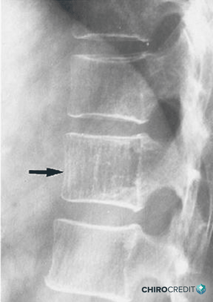
Note the coarsened vertical trabeculae producing a characteristic corduroy appearance at one level. Image from an upcoming course by Dr. Deltoff.
Reading the Film Is Only Half the Job
Knowing what to look for matters, but so does knowing what to order in the first place. Choosing the right imaging study for a given presentation — plain film versus advanced imaging, when to escalate, when an X-ray won’t answer the clinical question at all — is a distinct skill from interpretation, and it’s one that’s easy to under-invest in relative to the reading itself.
Building the Eye Systematically
This is where structured, radiologist-led continuing education earns its place. ChiroCredit’s Diagnostic Imaging library includes more than 40 courses taught by DACBR-credentialed instructors and MD/DC radiologists, covering everything from systematic approaches to reading plain film spine films, to chest radiology, to imaging safety and radiation protection, to the specific pathology patterns most likely to be missed by generalist review. It’s built for exactly the gap described above: not a single course, but a systematic way to sharpen the read over time.
More reads like this — explore the full library: ChiroCredit Diagnostic Imaging CE Courses
References
- ChiroCredit Diagnostic Imaging course catalog (Chiropractic Doctor category). ChiroCredit.com. https://www.chirocredit.com/category/Diagnostic_Imaging/Chiropractic_Doctor — Primary source for course titles, instructor credentials (DACBR, MD/DC radiologists), and topic coverage cited in this article, including courses on benign/malignant neoplasia differentiation, arthritides, age-specific fracture patterns, chest radiology, and imaging ordering guidance.
